
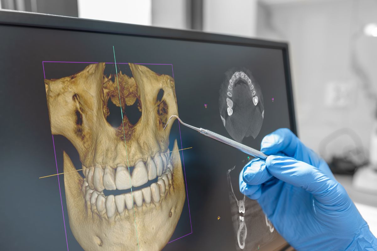
3D dental imaging, also known as Cone Beam Computed Tomography (CBCT), captures three-dimensional images of teeth, jaw, and facial structures. This advanced technology provides detailed volumetric data that allows for precise diagnostic assessment and surgical planning. 3D imaging is particularly valuable for complex cases involving implant placement, orthodontics, oral surgery, and temporomandibular joint assessment.
Advanced 3D imaging enables accurate assessment of bone density, implant site evaluation, and detailed surgical planning for complex procedures. 3D imaging reduces the need for multiple scans, provides superior visualization of anatomical structures, and improves clinical decision-making. This technology is essential for specialized treatments and ensures optimal outcomes in advanced dental procedures.
Please note: A written interpretation of an image, upon the client's request, is charged separately.
Advanced 3D Imaging Benefits
Both 2D and 3D imaging services offer comprehensive diagnostic capabilities that help in early detection of issues, allowing for prompt treatment and prevention of more serious conditions.

- Accurate diagnostic imaging
- Precise treatment planning
- Advanced visualization technology
- Minimal radiation exposure
- Quick and reliable results
Advanced 3D imaging enables accurate assessment of bone density, implant site evaluation, and detailed surgical planning for complex procedures. 3D imaging reduces the need for multiple scans, provides superior visualization of anatomical structures, and improves clinical decision-making. This technology is essential for specialized treatments and ensures optimal outcomes in advanced dental procedures.
Clinical Applications of 3D CBCT Imaging
Cone Beam Computed Tomography (CBCT) technology delivers unparalleled 3D visualization capabilities, revolutionizing modern dental diagnostics. Our advanced imaging systems capture comprehensive three-dimensional data in seconds, providing clinicians with critical diagnostic information for treatment planning across multiple specialties.
- Precise implant placement planning with bone density and volume assessment
- Comprehensive evaluation of bone quality and quantity for surgical procedures
- Advanced diagnosis of dental cysts and maxillary sinus pathology
- Complex extraction planning for impacted and retained teeth
- Detailed mandibular nerve positioning assessment to prevent complications
- Maxillary sinus floor evaluation for upper jaw surgical procedures
- Precise measurement of bone density for implant stability prediction
- Temporomandibular joint (TMJ) disorder diagnosis and treatment planning
- Comprehensive periodontal disease assessment and bone loss evaluation
- Root canal anatomy visualization for complex endodontic cases
- Orthodontic treatment planning with accurate tooth and bone positioning
- Orthognathic surgery planning for jaw repositioning procedures
- Impacted tooth localization and customized treatment approach
- Osteonecrosis detection and monitoring for early intervention
- Osteomyelitis diagnosis and treatment response evaluation
FAQs
Everything you need to know about dental diagnostic imaging
2D imaging provides flat X-ray images of teeth and jaw, while 3D imaging (CBCT) creates detailed three-dimensional images for complex cases requiring precise surgical planning.
Dental imaging uses minimal radiation. 2D X-rays use very low doses, and modern 3D imaging (CBCT) has been optimized to reduce radiation while maintaining image quality.
We recommend routine 2D imaging annually or as needed, while 3D imaging is typically recommended for specific diagnostic or surgical planning purposes when clinically indicated.
Dental X-rays are considered safe. The radiation exposure is minimal and well below harmful levels. We use modern equipment and protective measures to further minimize exposure.
Yes, we welcome previous imaging records. Bringing your prior X-rays helps us reduce unnecessary repeat imaging and allows for better comparison and continuity of care.
